MSK Ultrasound FoundationS Course
Home > Online Courses > Foundations Course
Save $100
$1295
Preview the first lesson from each module
Course Information
Instructor: Dr. Randy Moore
Lessons: 50+
Duration: 13h+
CME Credits: Earn up to 13 CME Credits
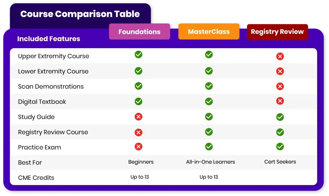
Course level: Beginners
Language: English

Lesson 50+
Students 700+
Beginner
MSK Ultrasound Fundamentals: Complete Training Course
Learn MSK ultrasound fundamentals with our comprehensive beginner training course. Build confidence in musculoskeletal sonography through 13 hours of expert-guided instruction, diagnosing with precision, and elevating your patient care.
Upper Extremity Course
Lower Extremity Course
Scan Demonstrations Course
Digital Textbook

Instructor:
Dr. Randy Moore
Category:
Musculoskeletal Sonography
Review:


Save $100
$1295
Preview the first lesson from each module
Course Information
Instructor: Dr. Randy Moore
Lessons: 50+
Duration: 13h+
CME Credits: Earn up to 13 CME Credits
Course level: Beginner to Expert
Language: English
GET STARTED
Take the next step in your career with the MSK Ultrasound Foundation Course — a concise, expert-led program that turns beginner curiosity into practical skill you can use the next day. Enroll today to gain hands-on scanning protocols, real-patient demonstrations, and a professional certificate with CME credits that immediately boosts your diagnostic confidence and practice value.

OVERVIEW
CURRICULUM
INSTRUCTOR
CME CREDITS
REVIEWS
GUARANTEE
FAQ
Begin LEARNING MSK Ultrasound
MSK Ultrasound Foundation Course Overview
Confidently elevate your diagnostic capabilities with the MSK Foundation Course—your fast track to mastering musculoskeletal ultrasound fundamentals. In just 13 hours, gain practical MSK ultrasound training skills that integrate seamlessly into your clinical workflow. Rapidly build competence in evaluating soft tissues, joints, and guided procedures through high-impact, expertly designed modules.
Professionally curated by pioneers in MSK sonography, this beginner-friendly course delivers a results-driven curriculum packed with clinical pearls and hands-on insights. Energize your medical practice with proven scanning protocols, HD scan demonstrations, and systematic MSK ultrasound training—led by one of the most respected voices in the field.
Revolutionize the way you visualize anatomy, interpret pathology, and guide treatments. Whether you're a physician, sonographer, or allied health professional new to MSK ultrasound, this foundation course empowers you to lead with clarity and confidence in musculoskeletal sonography.
What Will You Learn?
Whether you're new to MSK ultrasound or looking to build solid diagnostic fundamentals, this beginner course is built to deliver practical, high-yield learning. You'll move beyond theory and into actionable MSK ultrasound skills you can apply immediately in your clinical setting.
Develop systematic MSK ultrasound protocols for each joint and region
Master ultrasound anatomy of the upper and lower extremities
Identify common MSK pathologies with diagnostic confidence
Gain hands-on insight into ultrasound-guided injection procedures
Understand scanning techniques for soft tissue masses and spine imaging
Translate knowledge into clinical excellence through real-patient demonstrations
Included Courses
Whether you're new to MSK ultrasound or looking to sharpen your diagnostic edge, this foundation course is built to deliver practical, high-yield learning. You'll move beyond theory and into actionable skills you can apply immediately in your clinical setting.
Upper Extremity MSK Ultrasound Course
Shoulder, Elbow, Hand & Wrist
Build confidence in upper limb ultrasound with expert instruction in anatomy, pathology, and guided injection techniques. Covers MSK ultrasound imaging fundamentals, scanning protocols, and practical skills for real-world clinical use.
Lower Extremity MSK Ultrasound Course
Hip, Knee, Foot & Ankle
Build a strong foundation in lower limb ultrasound. Learn to identify normal and abnormal anatomy, recognize key pathologies, and perform accurate ultrasound-guided injections. Includes scanning protocols, clinical insights, and practical MSK ultrasound techniques you can apply right away.
MSK Ultrasound Scan Demonstrations Course
6 Regions: Shoulder, Elbow, Wrist, Hip, Knee, Foot & Ankle
Watch step-by-step scan demos with live narration by Dr. Randy Moore. Master real-time protocols and sonoanatomy on actual patients with split-screen guidance for hands-on MSK ultrasound learning.
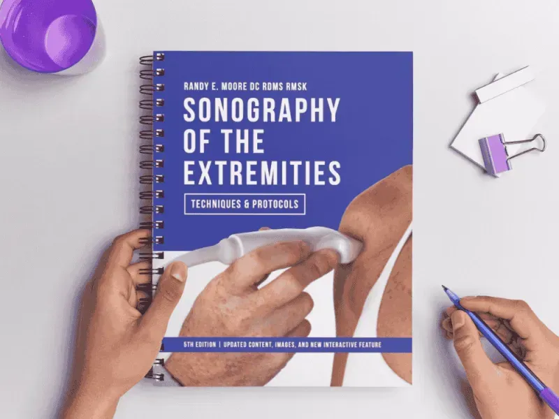
Textbook - NEW VERSION FOR 2026
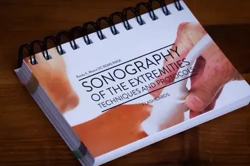
Sonography of the Extremities (5th Edition)
A comprehensive, step-by-step guide to MSK ultrasound protocols. Includes full-color images, interactive QR codes, labeled anatomy, positioning photos, scan tips, new modules and more.
Digital PDF Flipbook — Accessible anytime on phone, tablet, or desktop (online only).
Certification
Upon completion, receive a professional certificate that validates your proficiency in musculoskeletal ultrasound fundamentals. Earn CME credits and demonstrate your skills in clinical imaging, diagnosis, and procedural guidance. The program balances theoretical depth with real-world application—making it ideal for physicians, PAs, NPs, PTs, and sonographers beginning their MSK ultrasound journey.
Step-by-Step Training, All in One Place
Comprehensive Curriculum Overview
Our comprehensive MSK Ultrasound foundation curriculum is designed to take you step-by-step through essential anatomy, imaging fundamentals, and beginner scanning techniques across all major musculoskeletal regions. Each module combines expert guidance with practical demonstrations to help you build confidence in both diagnosis and ultrasound-guided interventions. Whether you're new to MSK ultrasound or seeking to establish solid fundamentals, this curriculum provides a structured, flexible learning path that fits your pace and goals.
Introduction & Handout
Get started with the course overview and essential materials to guide your MSK ultrasound learning journey.
Shoulder MSK Ultrasound: Complete Scanning Protocols
Explore detailed protocols and anatomy of the shoulder—the most mobile and injury-prone joint.
Elbow MSK Ultrasound: 360-Degree Assessment Techniques
Learn a 360-degree approach to elbow imaging for comprehensive assessment and diagnosis.
Hand & Wrist MSK Ultrasound: Complex Anatomy Scanning
Understand the complex anatomy and scanning techniques of the hand and wrist.
Hip MSK Ultrasound: Beyond Bursitis Diagnosis
Delve into hip imaging beyond bursitis, focusing on common pathologies and protocols.
Knee MSK Ultrasound: Comprehensive Above-to-Below Assessment
Cover the knee thoroughly—from above to below—for accurate diagnosis and evaluation.
Foot & Ankle MSK Ultrasound: Achilles and Plantar Fascia
Master scanning of the foot and ankle, including key structures like the Achilles tendon and plantar fascia.
Spine MSK Ultrasound: Lumbar Pathology Assessment
Focus on lumbar spine imaging to evaluate common back pathologies with precision.
Lower Extremity MSK Pathology Recognition
Recognize and diagnose a wide range of pathologies affecting the lower extremities.
Upper Extremity MSK Pathology Interpretation
Gain skills to identify and interpret abnormalities in the upper extremities.
Ultrasound-Guided Injection Procedures: Clinical Training
Learn safe and effective ultrasound-guided injection techniques for targeted treatment.

Meet Your Instructor
Randy E. Moore, DC, CMA, RDMS, RMSK
MSK Sonography Training | RMSK Exam Preparation | Clinical Ultrasound Education
Dr. Moore is a highly regarded and sought after pioneer, teacher and author in the quickly emerging field of musculoskeletal sonography. His straight forward, easily understood teaching methods have been deemed essential to shortening the learning curve for medical practitioners, medical sonographers, physician assistants, nurse practitioners, physical therapists and allied health professionals as they develop expertise in MSK sonography.
What Students Are Saying

Accredited & Trusted
Earn Up to 13 AMA PRA Catego24ry 1 Credits™
This activity has been planned and implemented in accordance with the accreditation requirements and policies of the Accreditation Council for Continuing Medical Education (ACCME) through the joint providership of Medical Academy, LLC, and MSK Masters. Medical Academy, LLC is accredited by the ACCME to provide continuing medical education for physicians.
Medical Academy, LLC designates this enduring activity for a maximum of 13 AMA PRA Category 1 credit(s)™.
Physicians should claim only the credit commensurate with the extent of their participation in the activity.

Risk-Free Enrollment
Enroll with confidence — backed by our 72-hour, no-questions-asked money-back guarantee.
We're so confident that you'll love MSK Masters and our one-of-a-kind training courses, that we offer a full 72 hour money back guarantee. If you don't find our online courses as informational and impactful as we say, then simply let us know and we'll refund your money.
Period.
Frequently Asked Questions
New to MSK ultrasound? Get quick answers to the most common questions about the Foundation Course.
Who is this course for?
This course is designed for medical professionals who are new to musculoskeletal ultrasound. It’s ideal for physicians, sonographers, PAs, NPs, PTs, and other allied health providers looking to build a strong diagnostic foundation.
Do I need any prior ultrasound experience?
No prior ultrasound experience is required. The course begins with the basics and builds progressively, giving you the structure and confidence to learn at your own pace.
How long do I have access to the course?
You get lifetime access to all course materials, including any future updates. Learn on your own schedule, revisit lessons anytime, and stay current with evolving best practices.
Is there a certificate of completion?
Yes. Upon completing the course, you’ll receive a professional certificate that verifies your proficiency in MSK ultrasound fundamentals.
Does this course offer CME credits?
Yes. The course is eligible for up to 13 AMA PRA Category 1 Credits™. These can be claimed after completing the curriculum and passing the assessment.
What’s included in the course?
You’ll get comprehensive training modules, HD scan demonstrations, region-by-region protocols, and the full Sonography of the Extremities (4th Edition) digital textbook. Everything is delivered in a beginner-friendly, step-by-step format.
Is there a refund policy?
Yes. We offer a 72-hour, no-questions-asked money-back guarantee. If you’re not satisfied for any reason, just contact us within three days of purchase for a full refund.
Your Sneak Peek Starts Here
Free Course Previews
Imaging Fundamentals I
A practical intro to MSK ultrasound focused on normal anatomy, image optimization, and essential scanning techniques.
The Shoulder I
Learn a step-by-step shoulder ultrasound protocol focusing on biceps and subscapularis tendon imaging. Covers patient positioning, probe techniques, and key anatomical landmarks for accurate, dynamic MSK shoulder evaluation.
The Elbow I
This video covers anterior elbow ultrasound, focusing on the distal biceps tendon. Learn probe positioning, patient setup, and key approaches—especially lateral and dorsal—for clear, accurate imaging of elbow anatomy.
The Hip I
This video covers anterior hip ultrasound, focusing on probe selection, patient positioning, and identifying key structures like the joint capsule and labrum. Includes tips for detecting effusion and performing safe, guided injections.


