Home > Blog
BLOG
Your Injection is No Better than Your Image: Raising the Bar in Regenerative Medicine Treatment
This statement is not meant to be confrontational in any sense of the word. It is, though, an irrefutable fact, and a call to maintain the highest standard of bio-cellular delivery. In a previous post, the direct relationship of optimal response to precise placement of the regenerative/bio-cellular product was emphasized. It’s true!
I have had the opportunity to develop MSK protocols in four regenerative practices. Without exception, the facilities reported vastly more positive response to treatment. Why? For example, in a shoulder case, MSK Ultrasound readily identifies bursal verses articular side supraspinatus defects . . . such as the degenerative changes of tendinosis. It is not always a tear!
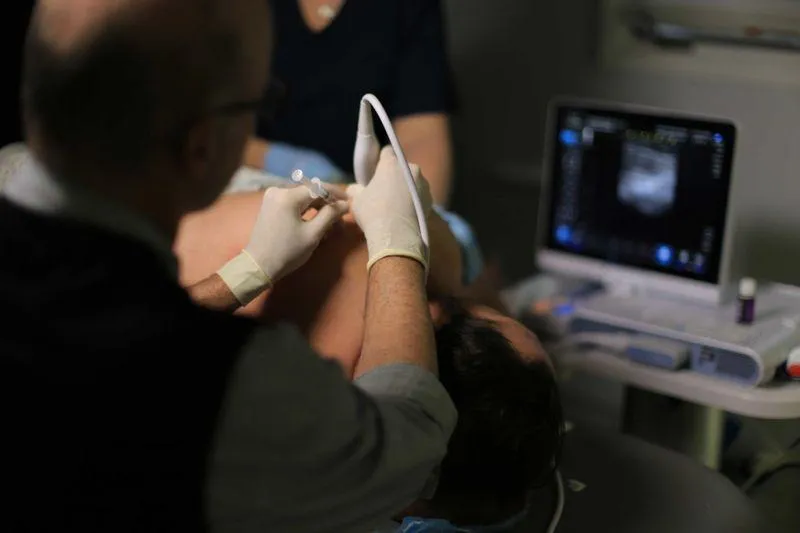
AC Joint Instability and/ or Separation of Dynamic Real-Time Imaging
I have found supraspinatus impingement under the acromion to be rare and AC Joint Impingement on a frequent basis. AC Joint instability responds very well to precise placement with fenestration of the of the ligament/capsule complex. Multiple Sites within the same tendon, ligament or muscle often require specific deposits of bio-cellular product. Full spectrum or comprehensive treatment of an extremity can be as many as 8-10 specific sites. Ultrasound is the only modality that can offer this amazing specificity of treatment.
Additional Frequent Sites of Shoulder Treatment
Rotator Cuff Interval: Plays a Prominent Role in shoulder stability
Subscapularius Tendon: Range of Motion restrictions with calcification of tendinosis
True Intra-Articular Glenohumeral injections avoiding the labrum.
Build a Firm Foundation of MSK Skills. MSK Masters presents a simple, straightforward learning model.
1. Image Generation: Standardized/Reproducible patient and probe positioning
2. Image Recognition: Standardized Interpretation Algorithm
3. Image Interpretation: Pathology identified via keen understanding of normal sonoanatomy.